详情
Overview:
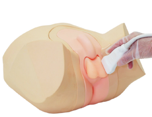
The Scrotal Ultrasound Training Model is designed based on real adult male pelvic data, accurately replicating anatomical structures such as the left and right testicles, epididymal head, epididymal body, and epididymal tail. The model material closely matches the acoustic properties of human tissue. The scrotum and its pathological features—such as size, orientation, and structure—are consistent with those of real adults. The model includes pathological conditions such as intratesticular masses, epididymal masses, and hydrocele, enabling the detection of both normal and pathological testicles.
Features:
The model material exhibits acoustic properties similar to human tissue, including density, sound velocity, and attenuation coefficient, making it compatible with ultrasound equipment of any brand.
One side features normal testicles and epididymis, while the other side presents pathological testicles and epididymis, facilitating comparative learning.
The anatomical structures are accurate, allowing for real ultrasound probe training on the model, including probe manipulation techniques and ultrasound image optimization.
The scrotal anatomy is precise, with scrotal and pathological features matching the size of real adults. Ultrasound can detect anatomical structures such as the left and right testicles, epididymal head, body, and tail, along with pathological conditions like intratesticular masses, epididymal masses, and hydrocele.
The model supports two-dimensional, three-dimensional, and four-dimensional ultrasound skill training.